Products
Equipment
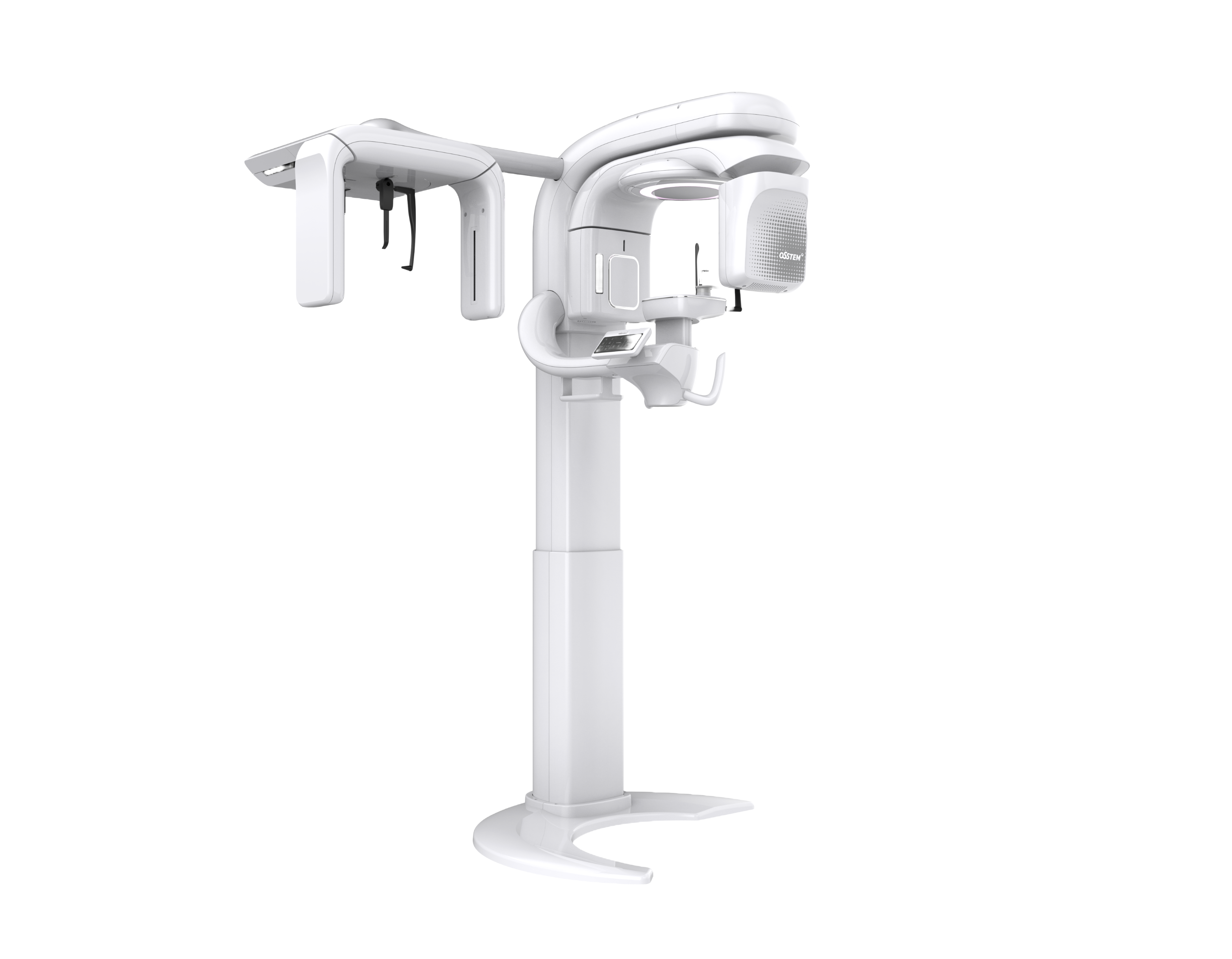
T2 CBCT
3 IN 1 Dental CBCT
Wide FOV, Clear Image and User-friendly CBCT
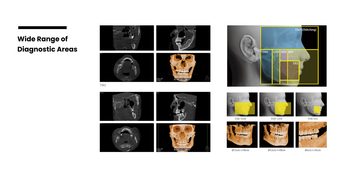
T2 offers six different imaging sizes with its wide Field of View (FOV). Its sizes range from 5x5, which produces clear images with an 80-voxel size, to 15x15, which allows for maxillofacial imaging. When combined with One3 software, a powerful 3D viewer, accurate diagnoses are just a click away. Experience the power of T2 CBCT.
Minimum FOV 15X15 enables Full-arch Imaging
T2 Stitching mode allows 15x15 imaging of orbital structures to the tip chin. This allows intraoral diagnoses, including TMJ and Sinus assessments.
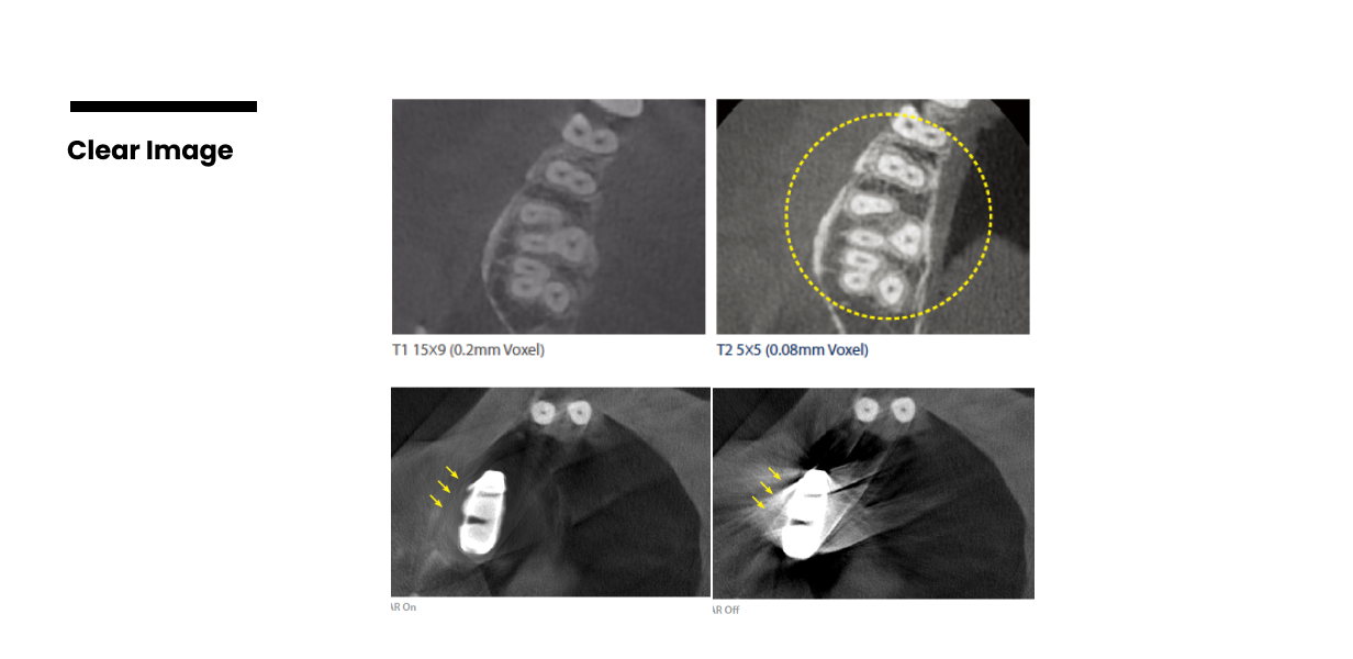
Clear Images
FOV 5X5 Endo-mode Imaging
A voxel is a 3D pixel, where the resolution of the anatomical structure increases with decreasing voxel size. In T2's high-definition FOV 5x5 endo-mode, images captured with 80μm voxel size allow for clear visualization of the alveolar bone and irregular root canals. This aids in precise endodontic therapy and leads to successful results.
Metal Artifact Reduction
T2's MAR function allows for the acquisition of clear images by automatically processing metal artifacts due to existing permanent restorations. This feature allows for accurate treatment plans based on distortion-free images, even in the oral environment of patients with existing metal fixed restorations.
Smart 3D Viewer, One3
The T2 comes equipped with 2D and 3D software as a standard feature. The T2's 3D software and viewer, named One3, provides the ability to perform complex tasks ranging from diagnosis to implant simulation on the multi-planner reconstruction (MPR) screen. The "Dental Arch" tab allows for planning, tracing and saving up to 5 dental arches, making it easy to analyze the cross-section of the images in the planned area.
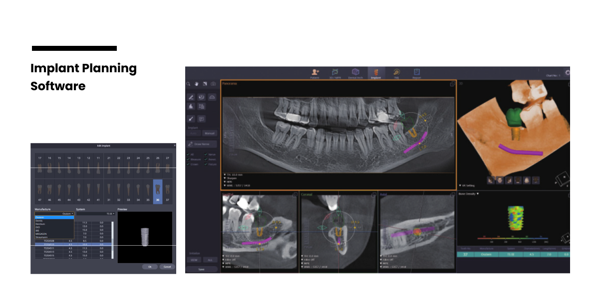
Optimizing Surgical Plans
One3 Implant Planning Mode
One3's implant planning mode enables the simulation of implant placement based on the actual design. The library includes a wide range of implants that can be simulated to match the design. Moreover, it facilitates the diagnosis of bone density around the surgical site, thereby enabling the planning of a safe and successful implant surgery.